- Home
- Editorial
- News
- Practice Guidelines
- Anesthesiology Guidelines
- Cancer Guidelines
- Cardiac Sciences Guidelines
- Critical Care Guidelines
- Dentistry Guidelines
- Dermatology Guidelines
- Diabetes and Endo Guidelines
- Diagnostics Guidelines
- ENT Guidelines
- Featured Practice Guidelines
- Gastroenterology Guidelines
- Geriatrics Guidelines
- Medicine Guidelines
- Nephrology Guidelines
- Neurosciences Guidelines
- Obs and Gynae Guidelines
- Ophthalmology Guidelines
- Orthopaedics Guidelines
- Paediatrics Guidelines
- Psychiatry Guidelines
- Pulmonology Guidelines
- Radiology Guidelines
- Surgery Guidelines
- Urology Guidelines
New ligament found in ankle

In a major breakthrough finding published in the journal Knee Surgery, Sports Traumatology, Arthroscopy, UB researchers defined a new anatomic structure in the ankle, the lateral tibiotalocalcaneal ligament complex (LFTCL) which could explain why many ankle injuries produce chronic pain.
Till now the anatomist knew that the ligaments in the ankle are grouped structured by two ligament complexes: the lateral collateral ligament -in the side of the joint and formed by three independent ligaments- and the medial or deltoid collateral ligament but the researchers found a new anatomic structure in the ankle named LFTCL.
Read Also: Management of ankle sprain-Updated guideline
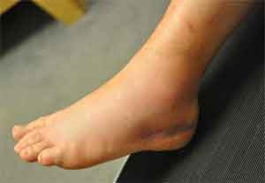
Ankle lateral ligaments are the ones that receive more injuries in the human body, especially due to ankle sprains. Moreover, many people who suffer from this injury complain about pain in the ankle which lingers and they are bound to get another sprain -which has not been explained in medicine yet.
"This lack of explanation was the key to change the way to tackle ligament dissection in the dissection room and we saw linking fibers between ligaments were usually removed because they were not associated with the ligament," says Miquel Dalmau Pastor, researcher from the Human Embryology and Anatomy Unit, and the Department of Pathology and Experimental Therapeutics of the UB.
Jordi Vega and associates performed the anatomical study in 32 fresh-frozen below-the-knee ankle specimens. A plane-per-plane anatomical dissection was performed. Overdissecting the area just distal to the inferior ATFL fascicle was avoided to not alter the original morphology of the ligaments and the connecting fibers between them. The characteristics of the ATFL and CFL, as well as any connecting fibers between them, were recorded. Measures were obtained in plantar and dorsal flexion, and by two different observers.
Read Also: Casting & surgery equally effective in ankle fractures in Elderly
The study found that ATFL was observed as a two-fascicle ligament in all the specimens. The superior ATFL fascicle was observed intra-articular in the ankle, in contrast to the inferior fascicle. The mean distance measured between superior ATFL fascicle insertions increases in plantar flexion (median 19.2 mm in plantar flexion, and 12.6 mm in dorsal flexion), while the same measures observed in the inferior ATFL fascicle does not vary (median 10.6 mm in plantar flexion, and 10.6 mm in dorsal flexion).
Further, the inferior ATFL fascicle was observed with a common fibular origin with the CFL. The CFL distance between insertions does not vary with ankle movement (median 20.1 mm in plantar flexion, and 19.9 mm in dorsal flexion, n.s.). The inferior ATFL fascicle and the CFL were connected by arciform fibers, that were observed as an intrinsic reinforcement of the subtalar joint capsule.
"Since the intra-articular ligament does not cicatrize, the instability of the joint produces pain so these patients are likely to suffer from another sprain and develop other injuries in the ankle," highlights Francesc Malagelada, the co-author of the study.
The study concluded that the superior fascicle of the ATFL is a distinct anatomical structure, whereas the inferior ATFL fascicle and the CFL share some features being both isometric ligaments, having a common fibular insertion, and being connected by arciform fibers, and forming a functional and anatomical entity, that has been named the lateral tibiotalocalcaneal ligament (LFTCL) complex.

Disclaimer: This site is primarily intended for healthcare professionals. Any content/information on this website does not replace the advice of medical and/or health professionals and should not be construed as medical/diagnostic advice/endorsement or prescription. Use of this site is subject to our terms of use, privacy policy, advertisement policy. © 2020 Minerva Medical Treatment Pvt Ltd