- Home
- Editorial
- News
- Practice Guidelines
- Anesthesiology Guidelines
- Cancer Guidelines
- Cardiac Sciences Guidelines
- Critical Care Guidelines
- Dentistry Guidelines
- Dermatology Guidelines
- Diabetes and Endo Guidelines
- Diagnostics Guidelines
- ENT Guidelines
- Featured Practice Guidelines
- Gastroenterology Guidelines
- Geriatrics Guidelines
- Medicine Guidelines
- Nephrology Guidelines
- Neurosciences Guidelines
- Obs and Gynae Guidelines
- Ophthalmology Guidelines
- Orthopaedics Guidelines
- Paediatrics Guidelines
- Psychiatry Guidelines
- Pulmonology Guidelines
- Radiology Guidelines
- Surgery Guidelines
- Urology Guidelines
Wilms tumour- GOI Standard Treatment Guidelines

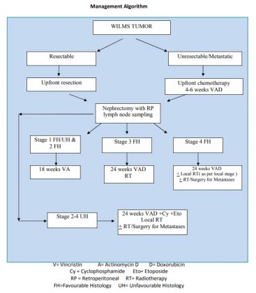
Dr Anil K. D’ Cruz, Director and chief Head and Neck Services, Tata Memorial Hospital, Mumbai has prepared Standard Treatment Guidelines for Wilms tumour on behalf of Ministry of Health and Family Welfare, Government of India which has released the same.
Wilms tumour is the most common renal malignancy in children. It predominantly affects children under 5 years of age, most commonly during the first 2 years of life. Contemporary treatment of Wilms tumour has led to an overall survival of over 85% and the emphasis is now shifting from successful treatment to reducing treatment-associated morbidity without loss of efficacy.
Following are the major recommendations :
Radiological report for Wilms Tumour
Should be a contrast enhanced CT/MRI of abdomen
Laterality: Right or Left or Bilateral disease
Location: which pole
Size
Local extent:
- Is it Completely Intra-renal location or is Perinephric spread evident
- Renal sinus infiltration.
- Renal vein status
- IVC status.
- Renal artery : comment about the abdominal origin as well, comment on accessory arteries if any
- Infiltration of liver, pancreas, bowel, GB, or abdominal wall
Significant ascites, Peritoneal implants
Adenopathy: Enlarged nodes
Metastatic disease: Lung, liver, bone or extra-abdominal nodes
Synoptic Pathology report for a renal tumour
Gross Examination: Received a specimen of right / left radical nephrectomy / nephrectomy/partial nephrectomy measuring -------X------X-------- cm. including perinephric fat. Hilum of the kidney shows ureter measuring --------cm along with arterial and venous stumps.
On cutting open, the kidney measures ----------X--------X--------cm and shows a tumour measuring--------X--------X-------- cm involving---------------------------------of the kidney. Cut surface of the tumour is solid / nodular solid / solid-cystic. Areas of necrosis and hemorrhage are-----------------------. Tumour invades through the renal capsule and extends into perinephric fat grossly / doesnot invade the renal capsule. Tumour involves / does not involve the Gerotta’s ( renal )fascia.
Renal sinus is----------------. Renal vein invasion is--------------------------------. Renal pelvis and ureter are-----------------------------. Resection end of ureter is ------------------------------- Resection end of renal vein is -----------------------------. Hilum of the kidney reveals-----------------nodes. Foci of nephroblastomatosis are not indentified / identified . A slice of the tumour is cut in a grid fashion and submitted for histopathology examination.
Microscopy:
Right / Left radical nephrectomy / Nephrectomy / Partial nephrectomy ,
Post chemotherapy / History of neo-adjuvent chemotherapy unknownTriphasic
/ Biphasic / blastemal predominant / teratoid Wilm’s tumour of ------------kidney.
-------------------------------------------------------------------------------------------------------------------------------
-------------------------------------------------------------------------------------------------------------------------------
----------------------------------------------------------------------
Favourable histology ( FH ) / unfavourable histology (UH )
Anaplasia is absent. Focal / diffuse anaplasia is seen.
Vascular invasion --------------------
Tumour does not invade the renal capsule / invades the renal capsule and is seen in perinephric fat..
Gerotta’s fascia is -----------------------------------
Renal sinus involvement is-------------------------------------
Renal vein involvement is ----------------------------
Hilar lymph nodes----------------------------------------------------------------------------------
Foci of nephroblastomatosis are not seen / -----------------type of nephroblastomatosis is present. -------------------------------------------------------------------------------------------------
Resection margins of ureter, renal vein and renal artery are ---------------------------------Adrenal is --------------------------------------------------------------------------------------------------------------------------------------------------------------------------------------------------------------------------------------------------------------------------------------------------------------------------------------------------------------------------------------------------------------------------------------------------------------------------------------------------------------------------------------------------------------------------------------------------------------------------------------------------------
Impression:
Right / Left radical nephrectomy / Nephrectomy / Partial nephrectomy ,
Post chemotherapy / History of neo-adjuvent chemotherapy unknown.:
Wilm’s tumour, ----------------histology, ---------------invading renal capsule / --------------------
invading Gerotta’s fascia, -- -------involving --------------lymph nodes.

For further reference log on to: http://clinicalestablishments.gov.in/WriteReadData/2611.pdf

Disclaimer: This site is primarily intended for healthcare professionals. Any content/information on this website does not replace the advice of medical and/or health professionals and should not be construed as medical/diagnostic advice/endorsement or prescription. Use of this site is subject to our terms of use, privacy policy, advertisement policy. © 2020 Minerva Medical Treatment Pvt Ltd