- Home
- Editorial
- News
- Practice Guidelines
- Anesthesiology Guidelines
- Cancer Guidelines
- Cardiac Sciences Guidelines
- Critical Care Guidelines
- Dentistry Guidelines
- Dermatology Guidelines
- Diabetes and Endo Guidelines
- Diagnostics Guidelines
- ENT Guidelines
- Featured Practice Guidelines
- Gastroenterology Guidelines
- Geriatrics Guidelines
- Medicine Guidelines
- Nephrology Guidelines
- Neurosciences Guidelines
- Obs and Gynae Guidelines
- Ophthalmology Guidelines
- Orthopaedics Guidelines
- Paediatrics Guidelines
- Psychiatry Guidelines
- Pulmonology Guidelines
- Radiology Guidelines
- Surgery Guidelines
- Urology Guidelines
Scientists find cause of facial widening defects

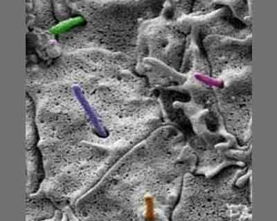
Widening across the forehead and nose occurs when loss of cilia at the surface of the cells disrupts internal signaling and causes two GLI proteins to stop repressing mid-facial growth. Ching-Fang Chang and Samantha Brugmann of the Cincinnati Children's Hospital Medical Center in Ohio, US, report the findings in a study published in PLOS Genetics.
Tiny projections, called cilia, are located on the surfaces of cells all over the body and serve an important role in cell signaling, which when lost, cause diverse diseases called ciliopathies. The mid-line of the face is especially susceptible to ciliopathies, and previous studies have shown that facial widening results from increases in Hedgehog signaling activity, a pathway that signals embryonic cells to direct development. However, the exact relationship between cilia loss and increased Hedgehog signaling is unclear. The scientists used mouse models of ciliopathies to investigate signaling proteins involved in this pathway, and looked specifically at the role of two proteins, GLI2 and GLI3. These proteins have both an activator form and a repress-or form that regulate expression of genes in the Hedgehog pathway. When they disrupted signaling from the cilia, the mice experienced facial widening because the cells lost the repress-or activity of GLI2 and GLI3 that normally keeps Hedgehog signaling in check.
The contributions of the activator and repress or functions of GLI genes to development is a complex topic that has been difficult for scientists to piece together. While the repress or forms of GLI3 and GLI2 were already known to play a role in neural tube and limb bud development, little was known about how these genes function in facial formation. These studies give insights into the mechanisms of how the cilia and GLI proteins function during mid-facial development.

Disclaimer: This site is primarily intended for healthcare professionals. Any content/information on this website does not replace the advice of medical and/or health professionals and should not be construed as medical/diagnostic advice/endorsement or prescription. Use of this site is subject to our terms of use, privacy policy, advertisement policy. © 2020 Minerva Medical Treatment Pvt Ltd