- Home
- Editorial
- News
- Practice Guidelines
- Anesthesiology Guidelines
- Cancer Guidelines
- Cardiac Sciences Guidelines
- Critical Care Guidelines
- Dentistry Guidelines
- Dermatology Guidelines
- Diabetes and Endo Guidelines
- Diagnostics Guidelines
- ENT Guidelines
- Featured Practice Guidelines
- Gastroenterology Guidelines
- Geriatrics Guidelines
- Medicine Guidelines
- Nephrology Guidelines
- Neurosciences Guidelines
- Obs and Gynae Guidelines
- Ophthalmology Guidelines
- Orthopaedics Guidelines
- Paediatrics Guidelines
- Psychiatry Guidelines
- Pulmonology Guidelines
- Radiology Guidelines
- Surgery Guidelines
- Urology Guidelines
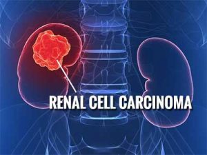
Renal Cell Carcinoma-Standard Treatment Guidelines

Renal cell carcinoma (RCC), which accounts for 2% to 3% of all adult malignant neoplasms, is the most lethal of the urologic cancers. The mortality rate of RCC is as much as twice that of bladder cancer1,2. There is no epidemiological data available from Indian subcontinent. However, the disease is fairly prevalent in our country. In 2010, an estimated 58,240 Americans were diagnosed with renal malignancies and 13,040 deaths were estimated3 . In 2008, there were an estimated 88,400 new cases and 39,300 kidney cancer–related deaths from RCC in Europe4.
Surgical excision remains the only curative treatment as this tumor is remarkably resistant to radiotherapy and chemotherapy.
Ministry of Health and Family Welfare, Government of India has issued the Standard Treatment Guidelines forRenal Cell Carcinoma.
Following are the major recommendations :
Presentation-
- Incidental: detected on imaging (CT / ultrasound) performed for other indication.
- Symptomatic: local / metastatic / paraneoplastic.
- Flank pain, hematuria, mass.
- Weight loss, fever, night sweats, recent onset hypertension, anemia / polycythemia.
- Cervical lymphadenopathy / non-reducing varicocoele / pathological fracture.
- others
Workup-
Any mass lesion detected on ultrasound needs further imaging.
- CT / MRI abdomen & pelvis/ MR Urography – both without and with contrast (if renal functions permissible). MRI specifically indicated if disease is infiltrating adjacent organs or IVC thrombus (triphasic multiplanar CT or high resolution color Doppler ultrasound optional for the latter).
- Blood investigations-Hemogram, kidney functions, alkaline phosphatase (ALP), calcium, albumin, lactate dehydrogenase (LDH).
- Chest X-ray-in all cases. Further imaging (CT scan) required only if clinically indicated or primary tumor locally advanced or lymph-nodes enlarged.
- Bone scan-only if clinically indicated (bone pain, raised ALP) or primary tumor locally advanced or lymph-nodes enlarged.
- PET CT-not routinely recommended in the workup for RCC. Has good specificity but low sensitivity in the evaluation of metastatic disease. Currently, it may be considered in case of equivocal findings on conventional imaging, where detection of metastatic disease will influence management decision.
- Biopsy / FNAC-not required in most cases. Acceptable in the following indications:
- Considering inflammatory mass / lymphoma / metastasis, vague Radiology, multiple masses, associated significant lymphadenopathy.
- Considering non-surgical therapy (e.g. cryotherapy, systemic therapy in case of metastatic disease) / active surveillance (small renal masses) / watchful waiting.
Staging (American Joint Committee on Cancer, TNM staging system for renal cancer, 7th ed, 2010)
| Primary tumor Tx Primary tumor cannot be assessed T0 No evidence of primary tumor T1a Tumor dia ≤ 4 cm confined to kidney T1b Tumor dia > 4-7cm confined to kidney T2a Tumor dia > 7-≤10cm confined to kidney T2b Tumor dia > 10cm confined to kidney T3a Tumor grossly extends into renal vein or its muscle-branches; tumor invading perirenal or sinus fat. T3b Tumor grossly extends into vena cava below diaphragm T3c Tumor grossly extends into vena cava above diaphragm or invades its wall T4 Tumor invades beyond Gerota’s fascia (including contiguous spread into ipsilateral adrenal) Regional lymph nodes Nx Regional lymph nodes cannot be assessed N0 No regional lymph node metastasis N1 Metastasis in single regional lymph node (s) Distant metastasis M0 No distant metastasis M1 Distant metastasis |
| Stage Grouping | |||
| Stage I | T1 | N0 | M0 |
| Stage II | T2 | N0 | M0 |
| Stage III | T1-2 T3 | N1 N0-1 | M0 M0 |
| Stage IV | T4 Any T | Any N Any N | M0 M1 |
Stage wise treatment-
In addition to clinical stage, patient’s performance status (ECOG5) should be taken into consideration before deciding the treatment options.
Stage I
Preferred – nephron-sparing surgery if technically feasible
Optional – radical nephrectomy*
Others
- Active surveillance
- Ablative therapies (cryotherapy, radiofrequency ablation, microwave thermotherapy, high frequency focussed ultrasound, etc.)
Adrenalectomy / lymphadenectomy: not indicated, unless grossly involved intraoperatively (staging changed)
- In patients with early stage RCC radical nephrectomy is justified when NSS is technically not feasible / the patient understands the other option (NSS) and opts for RN.
Stage II
Preferred – radical nephrectomy
Optional (imperative setting**) – nephron-sparing surgery
Adrenalectomy / lymphadenectomy: not indicated
- Imperative indications of NSS: solitary kidney, compromised function or reserve of contralateral kidney (chronic renal insufficiency, severe diabetes mellitus, severe hypertension), bilateral synchronous tumors, familial RCC.
Stage III
Preferred – radical nephrectomy, with tumor thrombectomy (if present)
Lymphadenectomy – may be performed for better staging. LND in patients with high-risk disease improves stage assessment and may prolong survival6,7. A mere sampling of the renal hilar lymph nodes is insufficient for pathologic staging. For right sided tumor, paracaval and interaortocaval lymph nodes and for left sided tumor para-aortic and interaortocaval lymph nodes should be removed from the crus of the diaphragm to the common iliac artery. If disease is confirmed within the interaortocaval nodes, a complete retroperitoneal LND is recommended to define the full extent of metastatic lymph node involvement.
Adrenalectomy: not required unless direct invasion or tumor nodule (stage changed).
Stage IV
- Nephrectomy with adjacent organ excision
- Metastasectomy (if solitary metastasis)
- Adrenalectomy
- Cyto-reductive nephrectomy
Patients of stage IV disease are candidates for adjuvant systemic therapy (vide infra).
Socio-economic and facility issues-
Advanced
- staging tools
- surgical facility
- follow up facility
- socio-economic support
may not be available everywhere.
Centers which intend to treat RCC must be equipped with facility for histopathology / CECT / Blood transfusion.
If above facilities not available / locally advanced disease / patient wants NSS → ref. to higher center.
Follow up protocol after definitive local treatment-
Risk grouping (UCLA integrated staging system)9:
| T stage | 1 | 2 | 3 | ||||||
| Furhman grade | 1-2 | 3-4 | 1-4 | 1 | 2-4 | ||||
| ECOG PS | 0 | ≥ 1 | ≥ 1 | Any | 0 | ≥ 1 | 0 | ||
| Risk group | Low | Intermediate | high | ||||||
Follow up protocol based on the above risk grouping10
| 6m | 12m | 18m | 24m | 30m | 36m | 42m | 48m | 54m | 60m | 6y | 7y | 8y | 9y | 10y | ||
| Hx/Ex/labs | L | - | √ | - | √ | - | √ | - | √ | - | √ | - | - | - | - | - |
| I | √ | √ | √ | √ | √ | √ | - | √ | - | √ | √ | √ | √ | √ | √ | |
| H | √ | √ | √ | √ | √ | √ | - | √ | - | √ | √ | √ | √ | √ | √ | |
| CT chest* | L | - | √ | - | √ | - | √ | - | √ | - | √ | - | - | - | - | - |
| I | √ | √ | √ | √ | √ | √ | - | √ | - | √ | √ | √ | √ | √ | √ | |
| H | √ | √ | √ | √ | √ | √ | - | √ | - | √ | √ | √ | √ | √ | √ | |
| CT abdomen | L | - | - | - | √ | - | - | - | √ | - | - | - | - | - | - | - |
| I | - | √ | - | √ | - | - | - | √ | - | - | √ | √ | √ | √ | √ | |
| H | √ | √ | √ | √ | - | √ | - | √ | - | √ | √ | √ | √ | √ | √ |
* CT chest can be replaced with CxR after initial 3 years.
L – low risk; I – intermediate risk; H – high risk
group. Systemic therapy-
Indications:
- Metastatic RCC with resectable disease (cytoreductive nephrectomy and metastatectomy should be done whenever feasible).
- Non-resectable Locally advanced / metastatic RCC
Agents-
| Name | |
| Sunitinib | |
| Sorafenib | |
| Pazopanib | |
| Bevacizumab + IFN-α | |
| Tamsirolimus | |
| Everolimus | |
| IFN-α | |
| High dose IL-2 |
Targeted therapy in the form of tyrosine kinase inhibitors (1-4) and m-TOR inhibitors (5-6) have become first line systemic treatment for management of metastatic RCC. A substantial improvement in progression-free survival and overall survival has been achieved in large randomized controlled trials, when compared to Interferon-α. These agents have also been found to be effective in non-clear cell RCC, which are typically resistant to cytokines and interferons. Sarcomatoid variant is associated with poor prognosis, and a modest response with doxorubicin & gemcitabine is observed.
Limitations-
- high cost and limited availability
- significant side-effects
- efficacy
Note : Targetted therapy for metastatic RCC/ locally advanced RCC should be decided by Urologist and given in supervision of a Urologist.
Guidelines by The Ministry of Health and Family Welfare :
Prof. Rajesh Ahlawat
Chairman,
Department of Urology & Kidney Transplant,
Medanta Medicity
Reviewed By:
Dr Anup Kumar Gupta Head of Department Department of Urology VMMC and Safdarjang Hospital, New Delhi

Disclaimer: This site is primarily intended for healthcare professionals. Any content/information on this website does not replace the advice of medical and/or health professionals and should not be construed as medical/diagnostic advice/endorsement or prescription. Use of this site is subject to our terms of use, privacy policy, advertisement policy. © 2020 Minerva Medical Treatment Pvt Ltd