- Home
- Editorial
- News
- Practice Guidelines
- Anesthesiology Guidelines
- Cancer Guidelines
- Cardiac Sciences Guidelines
- Critical Care Guidelines
- Dentistry Guidelines
- Dermatology Guidelines
- Diabetes and Endo Guidelines
- Diagnostics Guidelines
- ENT Guidelines
- Featured Practice Guidelines
- Gastroenterology Guidelines
- Geriatrics Guidelines
- Medicine Guidelines
- Nephrology Guidelines
- Neurosciences Guidelines
- Obs and Gynae Guidelines
- Ophthalmology Guidelines
- Orthopaedics Guidelines
- Paediatrics Guidelines
- Psychiatry Guidelines
- Pulmonology Guidelines
- Radiology Guidelines
- Surgery Guidelines
- Urology Guidelines
Rare case of giant solitary fibrous tumor of the adrenal gland in a child: a report

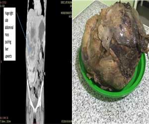
A 13-year-old Oromo girl presented with a progressively increasing right-sided abdominal mass, low-grade intermittent fever, and a dull right upper abdominal pain of 3 years’ duration with no other associated symptoms. There were no known past illnesses and there was no family history of similar illness. She was given pain medications and antibiotics on various occasions but there was no improvement.
Her general appearance was not acutely sick looking. Her vital signs were within normal limits. The pertinent abnormal finding was right-sided abdominal mass with well-defined medial and inferior border extending to right subcostal region. Complete blood count (CBC), urine analysis, and organ function tests were all normal. Ultrasound and a computed tomography (CT) scan demonstrated a huge vascular suprarenal mass displacing her right kidney caudally; its measurements were 16 × 19 cm and it contained multiple internal calcifications. There were no enlarged regional nodes and no vascular invasion.
Laboratory tests for functional adrenal tumors including serum and 24-hour urine metanephrines were all normal.
A working diagnosis of the huge nonfunctional adrenal tumor was made and our patient underwent exploratory surgery through a bilateral subcostal incision. The operative findings were a well-capsulated and highly vascularized mass arising from the superior aspect of her right kidney, which got a significant blood supply from the right lobe of the liver. The mass was successfully and completely resected and the specimen subjected to histopathology.
The histopathology report showed 18 × 15 × 12 cm white solid mass with necrotic center arising from the right adrenal gland. There was a patternless proliferation of spindle cells and ovoid cells that had mild pleomorphic nuclei and focally hyalinized stroma containing blood vessels. Mitosis was seen infrequently. These findings were consistent with an SFT of the adrenal gland.
Our patient was followed-up for 3 months and is doing remarkably well and members of her family were very grateful.
This is the sixth or seventh case of its kind in the world to the best of our knowledge.
For more details click on the link: doi: 10.1186/s13256-019-2163-z

Disclaimer: This site is primarily intended for healthcare professionals. Any content/information on this website does not replace the advice of medical and/or health professionals and should not be construed as medical/diagnostic advice/endorsement or prescription. Use of this site is subject to our terms of use, privacy policy, advertisement policy. © 2020 Minerva Medical Treatment Pvt Ltd