- Home
- Editorial
- News
- Practice Guidelines
- Anesthesiology Guidelines
- Cancer Guidelines
- Cardiac Sciences Guidelines
- Critical Care Guidelines
- Dentistry Guidelines
- Dermatology Guidelines
- Diabetes and Endo Guidelines
- Diagnostics Guidelines
- ENT Guidelines
- Featured Practice Guidelines
- Gastroenterology Guidelines
- Geriatrics Guidelines
- Medicine Guidelines
- Nephrology Guidelines
- Neurosciences Guidelines
- Obs and Gynae Guidelines
- Ophthalmology Guidelines
- Orthopaedics Guidelines
- Paediatrics Guidelines
- Psychiatry Guidelines
- Pulmonology Guidelines
- Radiology Guidelines
- Surgery Guidelines
- Urology Guidelines
PET Scans to Optimize Tuberculosis Meningitis Treatment, Study Finds

Despite treatment, over half of patients of Tuberculosis meningitisTBM die or have significant neurological injury lasting a lifetime, especially young children.
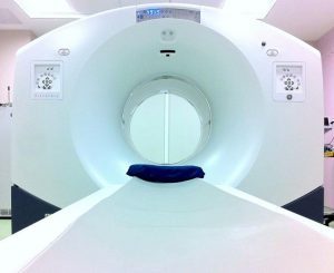
In the new study, Johns Hopkins researchers engineered a version of rifampin with a charged particle—called a positron—attached to the drug ([11C]rifampin) that allowed them to follow its movement throughout the body using PET (positron emission tomography) scans. This will go a long way to optimize and personalize the treatment of TBM. The findings of the new study have been published in Science Translational Medicine.
The findings, the researchers say, help advance understanding of why current drug treatments for TBM may not be as effective. And they say the PET approach offers a potential way to optimize the treatment of TBM, to ensure that enough drug is reaching the infected lesions.
“Really precise information has never been easy to come by for how much rifampin gets to any given patient where it’s needed,” says corresponding author Sanjay Jain, M.D., professor of paediatrics, radiology and international health at the Johns Hopkins University School of Medicine. “We’ve been able to use technology to find that long-needed information about this very troubling disease.”
Tuberculosis, which mostly infects the lungs, affects more than 10 million people around the world each year, causes more than 1 million deaths and costs the global economy billions of dollars, according to the World HealthOrganization. Tuberculosis meningitis (TBM) is often a deadly, always hard to treat, and a particular threat to young children. It may leave survivors with lifelong brain damage. Now, researchers at Johns Hopkins Medicine report they have used PET scans, a rabbit model and a specially tagged version of the TB drug rifampin to advance physicians’ understanding of this disease by showing precisely how little rifampin ever reach the sites of TB infection in the brain.
Jain, along with lead authors Elizabeth Tucker, M.D., assistant professor of anesthesiology and critical care medicine, and Alvaro Ordonez, M.D., research associate, pediatric infectious diseases at the Johns Hopkins University School of Medicine, as well as other Johns Hopkins and University of Maryland colleagues, published their findings Dec. 5 in Science Translational Medicine.
TBM caused when Mycobacterium tuberculosis infects brain tissue and the fluid surrounding the brain and spinal cord is considered the most lethal and disabling form of TB. Children under the age of 5, and those with chronic diseases—notably diabetes and HIV—are most likely to develop TBM. Like all TB diseases, TBM is treated with a combination of drugs, including isoniazid, rifampin, and pyrazinamide, taken for a year.
Because TBM symptoms are similar among rabbits and humans, the researchers created an experimentally infected colony of rabbits with TBM, injected them with the tagged drug and tracked levels of the tagged [11C]rifampin throughout the rabbits’ brains over six weeks. PET scans revealed that after two weeks of treatment, the penetration of [11C]rifampin into TBM brain lesions significantly decreased, from 32 percent to only 11 percent of the levels of the drug noted in the blood. Significantly, they say, the decrease was not reflected in samples of cerebrospinal fluid (CSF) taken from the rabbits, despite that CSF is currently used as a standard proxy for determining drug and infection levels in people.
“Until now, when treating patients, we’ve relied on a needle biopsy,” says Tucker. “We’ve assumed that what’s happening at the tip of the needle at one-time point in a sample is what’s happening throughout the affected organ.” The new results in the rabbit model, she says, suggest that it’s not that simple, at least in the animals. The penetration of rifampin varied among TBM lesions even within a single animal and changed over the course of the long treatment timeline.
The researchers also gave the tagged version of the drug to 10 adult human TB patients already receiving rifampin therapy. The patients were scanned at the Johns Hopkins PET Center in Baltimore, and these studies were performed under the Food and Drug Administration guidelines for new PET tracers. One of the patients, a 24-year-old female, had TBM. Similar to the results in rabbits, a PET scan showed that penetration of the drug into the patient’s brain lesions was limited to less than 5 per cent.
“If further research in animals and humans confirms that rifampin and other anti-TB drugs penetrate differently into their brains,” says Ordonez, “we foresee that imaging can help us figure out the concentration of drug needed and tailor the dose for individual patients.”
The team also expects that the rabbit model of TBM can be used to answer key questions about the disease and its treatment, including what baseline levels of antibiotics are required to penetrate brain lesions more fully.
“We used to think we could just give a drug and patients should get better,” says Jain. “Now, we have tools to measure the nuances that make a difference. PET could be used to study other antibiotics and also allow precision medicine approaches in resource-rich settings, such as the U.S. for other serious infections including MRSA (methicillin-resistant Staphylococcus aureus).”

Disclaimer: This site is primarily intended for healthcare professionals. Any content/information on this website does not replace the advice of medical and/or health professionals and should not be construed as medical/diagnostic advice/endorsement or prescription. Use of this site is subject to our terms of use, privacy policy, advertisement policy. © 2020 Minerva Medical Treatment Pvt Ltd