- Home
- Editorial
- News
- Practice Guidelines
- Anesthesiology Guidelines
- Cancer Guidelines
- Cardiac Sciences Guidelines
- Critical Care Guidelines
- Dentistry Guidelines
- Dermatology Guidelines
- Diabetes and Endo Guidelines
- Diagnostics Guidelines
- ENT Guidelines
- Featured Practice Guidelines
- Gastroenterology Guidelines
- Geriatrics Guidelines
- Medicine Guidelines
- Nephrology Guidelines
- Neurosciences Guidelines
- Obs and Gynae Guidelines
- Ophthalmology Guidelines
- Orthopaedics Guidelines
- Paediatrics Guidelines
- Psychiatry Guidelines
- Pulmonology Guidelines
- Radiology Guidelines
- Surgery Guidelines
- Urology Guidelines
Parathyroid carcinoma presenting as radial fracture

A unique and rare case appeared in NEJM which reported radial fracture due to parathyroid carcinoma. Taku Suzuki and Yumi Tomiie described the case of a 45-year-old man presented to the orthopaedic clinic with pain in his left forearm.
Parathyroid carcinoma (PC) is an uncommon disease that accounts for less than 2% of cases of primary hyperparathyroidism. PC is usually associated with a great variety of symptoms and severe laboratory changes, with calcium and PTH higher than those seen in adenomas. Parathyroid carcinoma is being reported as a tumour that induces primary hyperparathyroidism. It causes excessive secretion of the parathyroid hormone and increases the blood parathyroid hormone and calcium. .
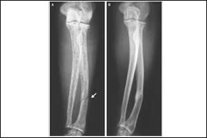
According to history, the patient had first felt the pain while gripping a railing 1 day earlier. A plain radiograph of the left arm showed a diaphyseal radial fracture (Panel A, arrow) and a diffusely low-density, mottled appearance of the radius and ulna.
Two months before presentation, the patient had received a diagnosis of primary hyperparathyroidism, with an intact parathyroid hormone level of 3590 pg per milliliter (reference range, 10 to 65) and a serum calcium level of 13.0 mg per deciliter (3.25 mmol per liter; reference range, 8.5 to 10.2 mg per deciliter [2.12 to 2.55 mmol per liter]).

Credit -NEJM Panel A Panel B
Courtesy NEJM
He underwent parathyroidectomy and was found to have the parathyroid carcinoma, which is a rare cause of primary hyperparathyroidism and can manifest as markedly elevated parathyroid hormone levels. Evaluation at the time of diagnosis showed no evidence of metastatic disease. The patient’s forearm was placed in a splint for 6 weeks, and he received vitamin D and calcium supplementation.
A plain radiograph obtained 1 year after the diagnosis of the fracture showed the union of the radial fracture and normal-appearing bone density (Panel B). One year after the parathyroidectomy, the patient’s parathyroid hormone level was 48 pg per millilitre and his calcium level was 9.0 mg per deciliter (2.25 mmol per litre).
In Parathyroid carcinoma, an increase of blood parathyroid hormone concentration due to hyperparathyroidism destroys the balance of osteoblasts and osteoclasts through activation of the osteoclast which may lead to pathological fracture.
For further reference log on to
DOI: 10.1056/NEJMicm1710426

Disclaimer: This site is primarily intended for healthcare professionals. Any content/information on this website does not replace the advice of medical and/or health professionals and should not be construed as medical/diagnostic advice/endorsement or prescription. Use of this site is subject to our terms of use, privacy policy, advertisement policy. © 2020 Minerva Medical Treatment Pvt Ltd