- Home
- Editorial
- News
- Practice Guidelines
- Anesthesiology Guidelines
- Cancer Guidelines
- Cardiac Sciences Guidelines
- Critical Care Guidelines
- Dentistry Guidelines
- Dermatology Guidelines
- Diabetes and Endo Guidelines
- Diagnostics Guidelines
- ENT Guidelines
- Featured Practice Guidelines
- Gastroenterology Guidelines
- Geriatrics Guidelines
- Medicine Guidelines
- Nephrology Guidelines
- Neurosciences Guidelines
- Obs and Gynae Guidelines
- Ophthalmology Guidelines
- Orthopaedics Guidelines
- Paediatrics Guidelines
- Psychiatry Guidelines
- Pulmonology Guidelines
- Radiology Guidelines
- Surgery Guidelines
- Urology Guidelines
Now parents can see their unborn babies in 3D VR models

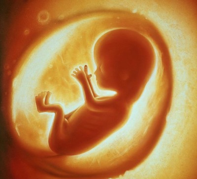
New York : In a new breakthrough research, Brazilian scientists have developed a new technology that will enable parents to watch their unborn babies grow in realistic three dimensional immersive visualisation.
The new technology combines magnetic resonance imaging (MRI) which provides high-resolution foetal and placental imaging with excellent contrast and ultrasound data to scan segments of the mother's womb and foetus to build a 3-D model which can be brought to life by using a virtual reality (VR) headset.
"The 3-D foetal models combined with virtual reality immersive technologies may improve our understanding of foetal anatomical characteristics and can be used for educational purposes and as a method for parents to visualise their unborn baby," said Heron Werner Jr. from the Clinica de Diagnostico por Imagem, in Rio de Janeiro, Brazil.
Sequentially-mounted MRI slices are used to begin construction of the model. A segmentation process follows in which the physician selects the body parts to be reconstructed in 3-D.
Once an accurate 3-D model is created including the womb, umbilical cord, placenta and foetus the virtual reality device can be programmed to incorporate the model, the study said.
The virtual reality foetal 3-D models are remarkably similar to the post-natal appearance of the newborn baby.
They recreate the entire internal structure of the foetus, including a detailed view of the respiratory tract, which can aid doctors in assessing abnormalities, Werner added.
The technology also can help coordinate care with multidisciplinary teams and provide better visual information to parents to help them understand malformations and treatment decisions.
"We believe that these images will help facilitate a multidisciplinary discussion about some pathologies in addition to bringing a new experience for parents when following the development of their unborn child," Werner said.
The study will be presented at the annual meeting of the Radiological Society of North America in Chicago, US.

Disclaimer: This site is primarily intended for healthcare professionals. Any content/information on this website does not replace the advice of medical and/or health professionals and should not be construed as medical/diagnostic advice/endorsement or prescription. Use of this site is subject to our terms of use, privacy policy, advertisement policy. © 2020 Minerva Medical Treatment Pvt Ltd