- Home
- Editorial
- News
- Practice Guidelines
- Anesthesiology Guidelines
- Cancer Guidelines
- Cardiac Sciences Guidelines
- Critical Care Guidelines
- Dentistry Guidelines
- Dermatology Guidelines
- Diabetes and Endo Guidelines
- Diagnostics Guidelines
- ENT Guidelines
- Featured Practice Guidelines
- Gastroenterology Guidelines
- Geriatrics Guidelines
- Medicine Guidelines
- Nephrology Guidelines
- Neurosciences Guidelines
- Obs and Gynae Guidelines
- Ophthalmology Guidelines
- Orthopaedics Guidelines
- Paediatrics Guidelines
- Psychiatry Guidelines
- Pulmonology Guidelines
- Radiology Guidelines
- Surgery Guidelines
- Urology Guidelines
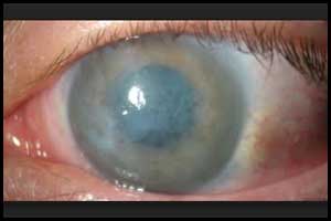
New treatment restores corneal clarity in patients with bullous keratopathy

Bullous keratopathy may be treated with an injection of cultured human corneal endothelial cells (CECs) supplemented with a Rho-associated protein kinase (ROCK) inhibitor, according to a study published in The New England Journal of Medicine.
Bullous keratopathy is a pathological condition in which small vesicles, or bullae, are formed in the cornea due to endothelial dysfunction.
Researchers of Japan conducted a study to investigate whether injection of cultured human CECs supplemented with a Rho-associated protein kinase (ROCK) inhibitor into the anterior chamber could increase CEC density. The study included eleven patients (avg age, 64.4 years) diagnosed with pseudophakic bullous keratopathy who had no detectable corneal endothelial cells. Each patient had a corneal thickness > 630 µm and visual acuity less than 20/40.
A total of 1×10⁶ passaged cells were supplemented with a ROCK inhibitor and injected into the anterior chamber of the eye that was selected for treatment. After the procedure, patients were placed in a prone position for 3 hours. The primary outcome was the restoration of corneal transparency, with a CEC density of more than 500 cells per square millimeter at the central cornea at 24 weeks after cell injection. Secondary outcomes were a corneal thickness of less than 630 μm and an improvement in best corrected visual acuity equivalent to two lines or more on a Landolt C eye chart at 24 weeks after cell injection.
The study found that all 11 eyes obtained a corneal endothelial cell density > 500 cells/mm2, with 10 eyes achieving 1000 cells/mm2 and six eyes exceeding 2000 cells/mm2at 24 weeks post-injection.
An improvement in both corneal structure and function was demonstrated, with a corneal thickness < 630 µm achieved in 10 of the 11 eyes and 82% of eyes having a two-line improvement in visual acuity. A sustained therapeutic effect was achieved; after 2 years, all 11 eyes maintained corneal transparency. The study found that CEC density of more than 500 cells per square millimeter was observed in 11 of the 11 treated eyes at 24 weeks. (100%), of which 10 had a CEC density exceeding 1000 cells per square millimeter.
Further, the results showed that a corneal thickness of fewer than 630 μm (range, 489 to 640) was attained in 10 of the 11 treated eyes, and an improvement in best corrected visual acuity of two lines or more was recorded in 9 of the 11 treated eyes.
The study concluded that injection of human CECs supplemented with a ROCK inhibitor was followed by an increase in CEC density after 24 weeks in 11 persons with bullous keratopathy.

Disclaimer: This site is primarily intended for healthcare professionals. Any content/information on this website does not replace the advice of medical and/or health professionals and should not be construed as medical/diagnostic advice/endorsement or prescription. Use of this site is subject to our terms of use, privacy policy, advertisement policy. © 2020 Minerva Medical Treatment Pvt Ltd