- Home
- Editorial
- News
- Practice Guidelines
- Anesthesiology Guidelines
- Cancer Guidelines
- Cardiac Sciences Guidelines
- Critical Care Guidelines
- Dentistry Guidelines
- Dermatology Guidelines
- Diabetes and Endo Guidelines
- Diagnostics Guidelines
- ENT Guidelines
- Featured Practice Guidelines
- Gastroenterology Guidelines
- Geriatrics Guidelines
- Medicine Guidelines
- Nephrology Guidelines
- Neurosciences Guidelines
- Obs and Gynae Guidelines
- Ophthalmology Guidelines
- Orthopaedics Guidelines
- Paediatrics Guidelines
- Psychiatry Guidelines
- Pulmonology Guidelines
- Radiology Guidelines
- Surgery Guidelines
- Urology Guidelines
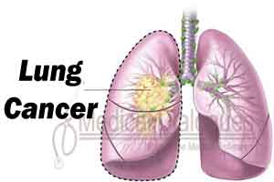
Lung Cancer - Standard Treatment Guidelines

Lung cancer is the commonest form of malignancy world over and is the most common cause of death. The disease is fatal if not treated early. Although, tremendous advances have taken place, the 5-year survival rate is still around 15%. Tobacco smoking is the most common cause of lung cancer and detection at the early stage is the key to successful treatment and prolonged survival.
Ministry of Health and Family Welfare, Government of India has issued the Standard Treatment Guidelines for Lung Cancer. Following are the major recommendations :
Case definition:
(For both situations of care)
It is a malignant condition of the lung and the tumor usually arises from the bronchi although this can originate from the alveoli (bronchoalveolar cell carcinoma). Bronchogenic carcinoma is of 4 major sub types: squamous cell carcinoma, adenocarcinoma, large cell carcinoma and small cell carcinoma. The first three are known as non small cell lung cancer.
Incidence of The Condition In Our Country
Lung cancer is the commonest form of malignancy in our country. It has surpassed the oropharyngeal carcinoma which used to be the commonest form. ICMR cancer registry, hospital based as well as community based, reveal it to be the commonest form of cancer in males and is amongst the top ten cancers in females. There is an increase in the incidence of lung cancer in recent years. The cancer atlas program also shows that the north eastern region of the country has the highest prevalence/incidence. It is estimated that every year nearly 58,000 new cases of lung cancer are diagnosed in the country.
Differential Diagnosis
- Tuberculosis
- Pneumonia
- Lung abscess
- Lung cysts
- Pleural effusion of any cause
Prevention And Counseling
Tobacco smoking cessation is the most important preventive measure. People working in high risk occupations with exposure to asbestos, arsenic, pollution, mercury, lead, and other heavy metals and chemicals need to have regular chest X-ray done.
Optimal diagnostic Criteria, Investigations, Treatment & Referral Criteria
*Situation 1: At Secondary Hospital: Optimal Standards of Treatment in Situations where technology and resources are limited
Clinical Diagnosis
Symptoms
Cough, expectoration, chest pain, haemoptysis, breathlessness, weight loss, anorexia, weakness, hoarseness of voice, dysphagia, superior vena cava (SVC) obstruction etc.
Signs
Clubbing, findings of collapse, mass lesion, haemorrhagic pleural effusion, hard lymph nodes and SVC obstruction are some of the common clinical findings.
Investigations
Chest radiograph: A chest radiograph is the cornerstone to establish the diagnosis of lung cancer which may reveal findings like mass, collapse, pleural effusion, or mediastinal lymph nodes. There may be consolidation particularly non-resolving. Bronchoalveolar cell carcinoma will have an alveolar pattern on chest radiograph.
Treatment:
Usually no treatment is possible at this level. The diagnosis need to be established first by other means as discussed below. However, the patients should be treated symptomatically with cough suppressants, pain relieving agents etc. Once the diagnosis and follow-up plan is made by a tertiary care hospital, the patient can be looked after for palliative therapy like pain management, thoracocentesis, cough suppressants and oxygen therapy etc.
Referral criteria:
1. Diagnosis not clear.
2. It is important that the doctor at this level should be able to suspect a diagnosis of lung cancer from the chest X-ray. It is common for these patients to be misdiagnosed as having tuberculosis and continue receiving anti-TB treatment and important time is lost before a diagnosis is achieved. Even if this is missed, a patient of tuberculosis should respond to ATT within a period of 2 to 3 weeks, failing which the patient should be referred.
*Situation 2: At Tertiary hospital where higher-end technology is available
Clinical Diagnosis
Symptoms
Cough, expectoration, chest pain, haemoptysis, breathlessness, weight loss, anorexia, weakness, hoarseness of voice, dysphagia, superior vena cava (SVC) obstruction etc.
Signs
Clubbing, findings of collapse, mass lesion, haemorrhagic pleural effusion, hard lymph nodes and SVC obstruction are some of the common clinical findings.
Investigations
- Chest radiograph: A chest radiograph is the cornerstone to establish the diagnosis of lung cancer which may reveal findings like mass, collapse, pleural effusion, or mediastinal lymph nodes. There may be consolidation particularly non-resolving. Bronchoalveolar cell carcinoma will have an alveolar pattern on chest radiograph.
- CT scan of chest (abdomen and brain is done for staging)
- Sputum cytology
- Fine needle aspiration cytology and biopsy
- Bronchoscopy
- Very rarely thoracotomy
- Mediastinoscopy (rarely done now)
- Bone scan (only in symptomatic patients)
- PET Scan (if available)
Treatment:
Out Patient
It is important to get a histological diagnosis and staging of the disease. The performance status (ECOG and Karnofsky scale) is to be ascertained. The overall financial conditions of the patient are to be accessed.
There are four treatment options:
- Early stage (Stage I to III A ) - surgery
- Stage III B and Stage IV -- chemotherapy
- Radiotherapy is usually a localized form of therapy and can be given in localized disease as a specific therapy. Both chemotherapy and radiotherapy can be given as an adjuvant therapy to surgery or as neo adjuvant setting to downstage the disease so that the patient is operable.
- Recently targeted therapy (molecular therapy) is available in certain types of lung cancers.
- Palliative or supportive therapy like pleurodesis in massive and repeated effusions, care of nutrition, pain alleviation, management of chemotherapy related toxicities like nausea and vomiting, diarrhea or constipation, hair loss etc. One also needs to look after the terminal care of the patient.
Suggested chemotherapy regimens:
A cisplatin based 2-drug combination therapy is recommended.
- cisplatin plus gemcitabine
- cisplatin plus docetaxel
- carboplatin plus paclitaxel
- cisplatin and paclitaxel
- cisplastin and irinotecan is a good and cheap combination therapy and can be used in our country.
Pemetrexed is a new anti-folate anti-metabolite useful in adenocarcinoma of the lung. It is to be given in combination with cisplatin. The above combinations is for non small cell lung cancer. For small cell lung cancer one can use cisplatin and irinotecan or etoposide.
Molecular therapy: epidermal growth factor receptor (EGFR) inhibitors like gefitinib and erlotinib are useful drugs for treating adenocarcinoma of the lung particularly in non-smoking and Southeast Asian female patients. Cetuximab is a monoclonal antibody also useful in these patients. Vascular endothelial growth factor receptor (VEGFR) inhibitors like bevacizumab are a new drug useful for non squamous cell type of non small cell lung cancer.
One should keep in mind that chemotherapeutic drugs are toxic and costly also. All the aspects should be explained to the patient as well as to the family before such a decision is taken.
WHO DOES WHAT? and TIMELINES
Doctor:
Usually the chest physician makes the diagnosis and the management should include a team of thoracic surgeon, medical oncologist and radiotherapist. However, a chest physician or an internist can handle such a case in delivering chemotherapy with proper training.
Nurse :
Implementation of orders, monitoring of patients and counseling, management of side-effects.
Technician:
Investigations
Resources Required For One Patient
| Situation | Human Resources | Investigations | Drugs & Consumable | Equipment |
| 1. | 1. Physician 2. Nurse 3. Radiographer | 1. Chest radiograph 2. Hemogram and blood chemistry | 1. Cough suppressant 2. Analgesics | 1. X-ray machine 2. Hematology and biochemistry |
| 2. | All above plus 1. ICU staff (Physician, nurses) 2. Radiologist 3. Pulmonologist 4. Pathologist 5. Oncologist 6. Thoracic surgeon 7. Anesthetist 8. Nuclear medicine specialist 9. Radiotherapist 10. Nurse and technician for assisting with bronchsocopy 11. Nurse and technician for assisting with interventional radiology procedures 12. Nursing staff trained in assisting thoracic surgery 13. Respiratory therapist | Above plus 1. Sputum cytology 2. FNAC cytology and biopsy 3. Bronchoscopy 4. Arterial blood gases 5. CT scan 6. Bone scan 7. PET scan (if available) 8. SPECT-CT to detect bone metastases | 1. Chemotherpay protocol (cisplatin, gemcitabine, docetaxel, paclitaxel, carboplatin) 2. Surgery protocol 3. Molecular therapy (gefitinib, erlotinib) 4. Radiotherapy | Above plus 1. Pathology laboratory (with facilities for special stains) 2. CT scan machine 3. ABG machine 4. Fiberoptic bronchoscope 5. Infusion pumps 6. PET scan machine (if available) 7. Bone scan machine 8. Operation theatre 9. Radiotherapy machine 10. SPECT-CT |
Guidelines by The Ministry of Health and Family Welfare :
Dr S.K. SHARMA AIIMS

Disclaimer: This site is primarily intended for healthcare professionals. Any content/information on this website does not replace the advice of medical and/or health professionals and should not be construed as medical/diagnostic advice/endorsement or prescription. Use of this site is subject to our terms of use, privacy policy, advertisement policy. © 2020 Minerva Medical Treatment Pvt Ltd