- Home
- Editorial
- News
- Practice Guidelines
- Anesthesiology Guidelines
- Cancer Guidelines
- Cardiac Sciences Guidelines
- Critical Care Guidelines
- Dentistry Guidelines
- Dermatology Guidelines
- Diabetes and Endo Guidelines
- Diagnostics Guidelines
- ENT Guidelines
- Featured Practice Guidelines
- Gastroenterology Guidelines
- Geriatrics Guidelines
- Medicine Guidelines
- Nephrology Guidelines
- Neurosciences Guidelines
- Obs and Gynae Guidelines
- Ophthalmology Guidelines
- Orthopaedics Guidelines
- Paediatrics Guidelines
- Psychiatry Guidelines
- Pulmonology Guidelines
- Radiology Guidelines
- Surgery Guidelines
- Urology Guidelines
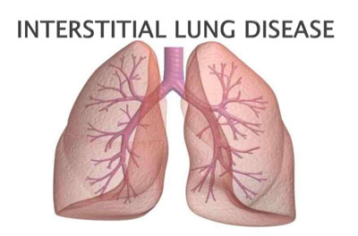
Interstitial Lung Disease - Standard Treatment Guidelines

Interstitial lung disease (ILD) is a heterogeneous group of disorders with a common clinico-radiological presentation, hence grouped together. These diseases are characterized by predominant involvement of lung ‘interstitium’ i.e. the space between alveolar basement membrane and pulmonary vascular endothelial membrane. As the disease progresses it encroaches alveolar spaces, terminal bronchioles and perivascular spaces, thereby ILDs are referred to as diffuse parenchymal lung disease (DPLD). ILDs due to systemic diseases such as connective tissue diseases, sarcoidosis, hypersensitivity pneumonitis, drugs, pneumoconioses and other miscellaneous conditions are termed as secondary ILDs. The primary pulmonary involvement is generally the Idiopathic pulmonary fibrosis (IPF/fibrosing alveolitis) or usual interstitial pneumonitis.
Ministry of Health and Family Welfare, Government of India has issued the Standard Treatment Guidelines for Interstitial Lung Disease. Following are the major recommendations :
Case definition:
For both situations of care:
ILD is suspected in patients with onset of respiratory symptoms of relatively acute/ sub-acute duration of weeks to months. The predominant symptoms consist of breathlessness and dry cough. Weakness and weight loss are usually present. Chest pain/ heaviness and haemoptysis can occur in a few conditions. Elicitation of occupational history is mandatory. The chest X-ray film shows distinctly reticular, reticulo-nodular or nodular opacities which are patchy /basilar or diffuse in distribution. Chest X-ray can occasionally be normal. High resolution CT scan of the chest is usually characteristic. Investigations for the underlying disease are important to rule out a secondary cause especially in those with atypical HRCT findings. Pathological diagnosis with bronchoscopic/ thoracoscopic and other surgical modalities is required for confirmation in only a few cases.
Incidence of The Condition In India
The exact incidence and prevalence of the disease is not known. The common causes of ILD in India include IPF, connective tissue related ILD (systemic sclerosis and rheumatoid arthritis) and sarcoidosis. In general, at most secondary- level hospitals, 1-3 cases per month and at super-specialty hospitals, 1-3 cases per week of idiopathic ILD are likely.
Differential Diagnosis
Many disorders can present with symptoms similar to that of ILD and interstitial opacities on chest radiograph. The following are the common disorders that should be ruled out before making a diagnosis of ILD:
- Tuberculosis
- Atypical pneumonias (Mycoplasma and legionella)
- Fungal pneumonias (Aspergillosis, crytococcosis)
- Chronic bronchitis
Exclusion of a secondary causes such as a connective tissue disease, hypersensitivity pneumonia, pneumoconiosis, drug-toxicity is important before making the diagnosis of primary idiopathic ILD (such as IPF).
Prevention And Counselling
Occupational and environment related interstitial lung disease may benefit from avoidance or decrease in intensity and duration of exposure. High index of suspicion and early recognition of interstitial lung disease can be helpful as it can prevent further deterioration of lung function by avoidance of exposure. Smoking cessation is another important caution which must be exercised.
The following are the few diseases in which avoidance of exposure can be advocated if ILD is detected early.
- Silicosis (occupations-miners, sandblasting, workers in abrasive industries, such as stone, clay, glass, cement manufacturing and granite quarrying)
- Asbestosis(asbestos manufacturing, construction trade, mining, milling and pipefitters)
- Coal worker’s pneumoconiosis
- Chronic hypersensitivity pneumonitis (pigeon breeders, farm dust and grain husk containing thermophilic actinomyces)
Optimal Diagnostic Criteria, Investigations, Treatment & Referral Criteria
While ILD can be suspected at any secondary-level hospital, the confirmation, exclusion of secondary causes and initiation of management lies essentially in the domain of the specialty hospitals. Follow-up management afterwards can, however, continue at the secondary –level, referring hospital.
Diagnosis:
The diagnosis of ILD is based on the combination of clinical, radiographic, and histopathology criteria.
- Clinical:
- Progressive breathlessness and/ or dry cough,
- Physical findings: basal end-inspiratory crackles- superficial, dry and velcro-like. Finger clubbing may be present in IPF.
- Radiographic findings: A good quality postero-anterior chest radiograph should be initial investigation of choice followed by HRCT chest for confirmation of presence of ILD.
- Chest radiograph- presence of reticular, nodular or reticulonodular shadows, shrunken lung fields and obscuration of cardiac and diaphragmatic margins.
- HRCT Chest- presence of reticular shadows (inter and intralobular septal thickening), patchy ground-glass opacities/consolidation (predominantly subpleural and peripheral) and honeycombing (indicates end-stage ILD)
- Pulmonary function tests:
- Spirometry : A restrictive abnormality is classical of ILD with reduced total lung capacity (TLC), functional residual capacity, and residual volume. Forced expiratory volume in one second (FEV1) and forced vital capacity (FVC) are reduced. FEV1/FVC ratio is normal or increased. However, mixed (restrictiveobstructive)/obstructive pattern can be seen in patients with lymphangioleiomyomatosis, sarcoidosis, hypersensitivity pneumonitis due to involvement of small airways.
- Diffusing capacity for carbon monoxide (DLCO) : reduced in all patients with ILD except in alveolar hemorrhage syndromes in which it is increased.
- Significant oxygen desaturation on exercise (>3-4%).
- ECG and 2D ECHO for pulmonary hypertension/cor pulmonale.
- Histopathology : The histopathology is required only in the presence of an early disease when the differential diagnosis is important from a treatable condition. The pattern varies according to the disease. Idiopathic pulmonary fibrosis is characterized UIP pattern. Sarcoidosis, Wegener’s granulomatosis and ChurgStrauss syndrome are characterized by granulomatous inflammation.
- 6-minute walk test (6MWT): For baseline and follow-up
Treatment :
The management of the patients with ILD is difficult and unsatisfactory. Antiinflammatory therapy with steroids, azathioprine or cyclophosphamide forms the main backbone of drug therapy. Interstitial lung diseases which respond well to immunosuppresive therapy include proliferative phase of NSIP, desquamative IP, cryptogenic organizing pneumonia, alveolar hemorrhage syndromes and sarcoidosis. Antioxidant and anti-fibrotic drugs such as N-acetyl cysteine and pirfenidone have some role for the treatment of idiopathic pulmonary fibrosis. None of the drugs are likely to provide benefit in advanced disease when only symptomatic treatment, home oxygen therapy, treatment of pulmonary hypertension, pulmonary rehabilitation and treatment of co-morbid diseases should be offered to improve quality of life and avoid unnecessary side-effects of the drugs.
Referral Criteria :
All patients in whom an interstitial lung disease is suspected or patients in whom alternative diagnosis like infection cannot be ruled out should be referred to a specialty hospital to confirm a diagnosis, identify the cause and initiate appropriate therapy.
WHO DOES WHAT?
Doctor
Evaluate the patients, order investigations, make a diagnosis, advise proper treatment and perform follow-up
Nurse
Carry out the Investigations suggested by the doctors and help in follow-up of patients
Technical staff
Perform relevant investigations as per the advice of the treating physician
Resources Required For One Patient
| Situation | Human Resources | Investigations | Drugs & Consumables | Equipment |
| 1. | 1.Physician 2. Nurse 3. Laboratory technician 4. Pulmonary function test technician 5. Radiographer 6. Physician with training in echocardiography / cardiologist | 1.Chest radiograph 2. Pulmonary function tests 3. ABG analysis 4. 2D-ECHO | 1.Parenteral and oral steroids (hydrocortisone, prednisolone) 2. N-acetyl cysteine 3. Inhaled bronchodilators 4. Sildenafil, bosentan | 1.Oxygen cylinder 2. Oxygen concentrators 3. Spirometer 4. Hand-held Spirometer 5. X-ray machine 6. ABG analyzer 7. ECHO machine |
| 2. | Above plus 1. ICU staff with pulmonary training 2. Radiologist 3. Pathologist 4. Physiotherapist | Above plus 1. CT Scan 2. Bronchoscopic biopsy (Transbronchial lung biopsy) | Above plus 1. Immunosuppressant drugs (azathioprine, cyclophosphamide, pirfenidone) | Above plus 1. ICU 2. Noninvasive and invasive ventilators 3. CT scan machine 4. Bronchoscope along with required facility |
Guidelines by The Ministry of Health and Family Welfare :
Dr S.K. SHARMA AIIMS

Disclaimer: This site is primarily intended for healthcare professionals. Any content/information on this website does not replace the advice of medical and/or health professionals and should not be construed as medical/diagnostic advice/endorsement or prescription. Use of this site is subject to our terms of use, privacy policy, advertisement policy. © 2020 Minerva Medical Treatment Pvt Ltd