- Home
- Editorial
- News
- Practice Guidelines
- Anesthesiology Guidelines
- Cancer Guidelines
- Cardiac Sciences Guidelines
- Critical Care Guidelines
- Dentistry Guidelines
- Dermatology Guidelines
- Diabetes and Endo Guidelines
- Diagnostics Guidelines
- ENT Guidelines
- Featured Practice Guidelines
- Gastroenterology Guidelines
- Geriatrics Guidelines
- Medicine Guidelines
- Nephrology Guidelines
- Neurosciences Guidelines
- Obs and Gynae Guidelines
- Ophthalmology Guidelines
- Orthopaedics Guidelines
- Paediatrics Guidelines
- Psychiatry Guidelines
- Pulmonology Guidelines
- Radiology Guidelines
- Surgery Guidelines
- Urology Guidelines
Hydropneumothorax in man with history of alcohol abuse associated cirrhosis

A case report published in The New England Journal of Medicine describes the case of a 47-year-old man presented with shortness of breath for the last 2 days which later turned out be hydropneumothorax on examination through chest radiography. The men had a history of cirrhosis associated with alcohol abuse.
"A 47-year-old man with a history of cirrhosis associated with alcohol abuse presented with a 2-day history of shortness of breath. Before this symptom developed, he had been treated with repeated thoracentesis of the right side for cirrhosis-associated hydrothorax," report Ping-Hsien Chen, and Xi-Zhang Lin from National Cheng Kung University, Tainan, Taiwan.
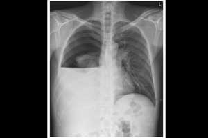
Hydropneumothorax is defined as the presence of both air (pneumothorax) and fluid (hydrothorax) within the pleural space. An upright chest x-ray will show air fluid levels.
On pulmonary examination, breath sounds were absent on the right side, and a succussion splash was audible in the right upper chest when the patient was gently shaken. Chest radiography showed hydropneumothorax with a collapsed right lung and an adjacent thoracic air-liquid level, which was probably the result of repeated thoracentesis.
The patient was treated with chest-tube placement and diuretics. An analysis of the pleural effusion revealed transudative fluid without evidence of infection or cancer. The chest drain was removed 1 week later, after reexpansion of the lung.
For more details click on the link: DOI: 10.1056/NEJMicm0810434

Disclaimer: This site is primarily intended for healthcare professionals. Any content/information on this website does not replace the advice of medical and/or health professionals and should not be construed as medical/diagnostic advice/endorsement or prescription. Use of this site is subject to our terms of use, privacy policy, advertisement policy. © 2020 Minerva Medical Treatment Pvt Ltd