- Home
- Editorial
- News
- Practice Guidelines
- Anesthesiology Guidelines
- Cancer Guidelines
- Cardiac Sciences Guidelines
- Critical Care Guidelines
- Dentistry Guidelines
- Dermatology Guidelines
- Diabetes and Endo Guidelines
- Diagnostics Guidelines
- ENT Guidelines
- Featured Practice Guidelines
- Gastroenterology Guidelines
- Geriatrics Guidelines
- Medicine Guidelines
- Nephrology Guidelines
- Neurosciences Guidelines
- Obs and Gynae Guidelines
- Ophthalmology Guidelines
- Orthopaedics Guidelines
- Paediatrics Guidelines
- Psychiatry Guidelines
- Pulmonology Guidelines
- Radiology Guidelines
- Surgery Guidelines
- Urology Guidelines
Horseshoe kidney in a deceased organ donor, a rare glimpse! LANCET case report

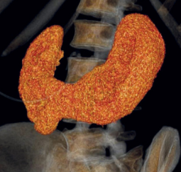
A very surprising and unique case of its kind was presented where a CT scan report of a man diagnosed with brain death showed a horseshoe kidney. The kidneys were fused at their lower poles with the isthmus overlying the aorta. The CT scan was done prior to the recovery of organs for donation. The case was reported in the journal The Lancet by Kamilia Butler-Peres and his associates.
A 42-year-old man was admitted to the trauma service with a single gunshot wound to the head. Upon diagnosis of brain death, he was referred for organ donation. The creatinine concentration was 1·00 mg/dL (estimated glomerular filtration rate [eGFR] 92·4 mL/min per 1·73 m2) and the urine output was more than 100 mL/h. Prior to recovery of organs for donation, the CT scan showed a horseshoe kidney. The kidneys were fused at their lower poles with the isthmus overlying the aorta. Hepato-pancreatic anatomy was only notable for a replaced right hepatic artery.
A 65-year-old woman with poorly controlled type 1 diabetes mellitus had been on hemodialysis for 4 months. She was offered a kidney from the above-cited donor by the transplant team. Each half of the donor's kidney had four arteries (two main renal arteries arose from the aorta and two arteries arose from the common iliac). However, each half of the donor's kidney had a single renal vein and ureter.
A vascular stapler was used to divide the renal isthmus in situ. The horseshoe kidney was divided in two prior to cross-clamp, allowing the transplant team to determine if the staple lines were hemostatic in the donor, rather than waiting until the organ was reperfused in the recipient.
The recipient had immediate graft function of the transplanted organ and was discharged with a creatinine concentration of 0·98 mg/dL (eGFR 69·3 mL/min per 1·73 m2). The mate kidney was transplanted into a separate recipient, and it also demonstrated immediate graft function.
For more reference log on to
https://www.thelancet.com/journals/lancet/article/PIIS0140-6736(18)30759-1/fulltext

Disclaimer: This site is primarily intended for healthcare professionals. Any content/information on this website does not replace the advice of medical and/or health professionals and should not be construed as medical/diagnostic advice/endorsement or prescription. Use of this site is subject to our terms of use, privacy policy, advertisement policy. © 2020 Minerva Medical Treatment Pvt Ltd