- Home
- Editorial
- News
- Practice Guidelines
- Anesthesiology Guidelines
- Cancer Guidelines
- Cardiac Sciences Guidelines
- Critical Care Guidelines
- Dentistry Guidelines
- Dermatology Guidelines
- Diabetes and Endo Guidelines
- Diagnostics Guidelines
- ENT Guidelines
- Featured Practice Guidelines
- Gastroenterology Guidelines
- Geriatrics Guidelines
- Medicine Guidelines
- Nephrology Guidelines
- Neurosciences Guidelines
- Obs and Gynae Guidelines
- Ophthalmology Guidelines
- Orthopaedics Guidelines
- Paediatrics Guidelines
- Psychiatry Guidelines
- Pulmonology Guidelines
- Radiology Guidelines
- Surgery Guidelines
- Urology Guidelines
Haemolytic Uremic Syndrome Revisited

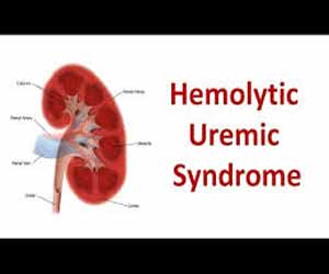
Hemolytic-uremic syndrome (HUS) is a group of blood disorders characterized by low red blood cells, acute kidney failure, and low platelets. Triad of clinical findings of HUS includes acute renal failure with oliguria, anuria, and rarely polyuria. Acute hemolytic anemia: microangiopathic with fragmented red blood cells (RBCs) or schistocytes, nonimmune, Coombs negative and thrombocytopenia. About 1.5 per 100,000 people are affected per year. Less than 5% of those with the condition die. Of the remainder, up to 25% have ongoing kidney problems. HUS was first defined as a syndrome in 1955.
CAUSES
Shiga toxin–producing E. coli OH157:H7 is the most frequent cause of HUS in the United States. Shiga toxin–producing E. coli O104:H4 infection caused a severe epidemic of HUS in Europe in 2011. Endothelial cell injury with secondary glomerular capillary microthrombi is central to the pathogenesis of HUS caused by Shiga toxin–producing E. coli. This is the most frequent cause of HUS, but there are other known causes of which atypical hemolytic uremic syndrome (aHUS) comprises about 10% of all causes. Genetic mutations increase the risk of aHUS and may lead to uncontrolled activation of the complement system when it is triggered.
Signs and symptoms
After eating contaminated food, the first symptoms of infection can emerge anywhere from 1 to 10 days later, but usually after 3 to 4 days. These early symptoms can include diarrhea (which is often bloody), stomach cramps, mild fever,or vomiting that results in dehydration and reduced urine. HUS typically develops about 5–10 days after the first symptoms, but can take up to 3 weeks to manifest, and occurs at a time when the diarrhea is improving. Related symptoms and signs include lethargy, decreased urine output, blood in the urine, kidney failure, low platelets, (which are needed for blood clotting), and destruction of red blood cells (microangiopathic hemolytic anemia). High blood pressure, jaundice (a yellow tinge in skin and the whites of the eyes), seizures, and bleeding into the skin can also occur. In some cases, there are prominent neurologic changes.
People with HUS commonly exhibit the symptoms of thrombotic microangiopathy (TMA), which can include abdominal pain, low platelet count, elevated lactate dehydrogenase LDH, a chemical released from damaged cells, and which is, therefore, a marker of cellular damage) decreased haptoglobin (indicative of the breakdown of red blood cells) anemia (low red blood cell count), schistocytes (damaged red blood cells),elevated creatinine (a protein waste product generated by muscle metabolism and eliminated renally, proteinuria (indicative of kidney injury), confusion, fatigue, swelling, nausea/vomiting, and diarrhea. Additionally, patients with aHUS typically present with an abrupt onset of systemic signs and symptoms such as acute kidney failure, hypertension (high blood pressure),myocardial infarction (heart attack), stroke,lung complications, pancreatitis (inflammation of the pancreas), liver necrosis (death of liver cells or tissue), encephalopathy (brain dysfunction), seizure, and coma. Failure of neurologic, cardiac, renal, and gastrointestinal (GI) organs, as well as death, can occur unpredictably at any time, either very quickly or following prolonged symptomatic or asymptomatic disease progression.
DIAGNOSIS
The similarities between HUS, aHUS, and TTP make differential diagnosis essential. All three of these systemic TMA-causing diseases are characterized by thrombocytopenia and microangiopathic hemolysis, plus one or more of the following: neurological symptoms (e.g., confusion, cerebral convulsions, seizures); renal impairment (e.g., elevated creatinine, decreased estimated glomerular filtration rate [eGFR], abnormal urinalysis); and gastrointestinal (GI) symptoms (e.g., diarrhea, nausea/vomiting, abdominal pain,gastroenteritis).The presence of diarrhea does not exclude aHUS as the cause of TMA, as 28% of patients with aHUS present with diarrhea and/or gastroenteritis. First diagnosis of aHUS is often made in the context of an initial, complement-triggering infection, and Shiga-toxin has also been implicated as a trigger that identifies patients with aHUS. Additionally, in one study, mutations of genes encoding several complement regulatory proteins were detected in 8 of 36 (22%) patients diagnosed with STEC-HUS. However, the absence of an identified complement regulatory gene mutation does not preclude aHUS as the cause of the TMA, as approximately 50% of patients with aHUS lack an identifiable mutation in complement regulatory genes.
Diagnostic work-up supports the differential diagnosis of TMA-causing diseases. A positive Shiga-toxin/EHEC test confirms a cause for STEC-HUS, and severe ADAMTS13 deficiency (i.e., ≤5% of normal ADAMTS13 levels) confirms a diagnosis of TTP.
TREATMENT
A study showed that children who received antibiotics (usually sulfa-containing or β-lactam antibiotics) during outbreaks had a higher rate (50% versus 7%) of HUS. A subsequent meta-analysis showed no protection and no association. Most specialists opt not to treat patients with E. coli OH157:H7 gastroenteritis with antibiotics because no benefit has been proven. Additional therapies, such as plasma exchange, immunoadsorption, Shiga toxin–binding agents and complement inhibitors (e.g., eculizumab).
Dr Gunjan Makkar is a columnist with medical dialogues and specialises in paediatric updates.

Disclaimer: This site is primarily intended for healthcare professionals. Any content/information on this website does not replace the advice of medical and/or health professionals and should not be construed as medical/diagnostic advice/endorsement or prescription. Use of this site is subject to our terms of use, privacy policy, advertisement policy. © 2020 Minerva Medical Treatment Pvt Ltd