- Home
- Editorial
- News
- Practice Guidelines
- Anesthesiology Guidelines
- Cancer Guidelines
- Cardiac Sciences Guidelines
- Critical Care Guidelines
- Dentistry Guidelines
- Dermatology Guidelines
- Diabetes and Endo Guidelines
- Diagnostics Guidelines
- ENT Guidelines
- Featured Practice Guidelines
- Gastroenterology Guidelines
- Geriatrics Guidelines
- Medicine Guidelines
- Nephrology Guidelines
- Neurosciences Guidelines
- Obs and Gynae Guidelines
- Ophthalmology Guidelines
- Orthopaedics Guidelines
- Paediatrics Guidelines
- Psychiatry Guidelines
- Pulmonology Guidelines
- Radiology Guidelines
- Surgery Guidelines
- Urology Guidelines
First-ever MRI waistcoat shall improve breast cancer screening

First-ever MRI waistcoat shall soon detect breast cancer in a more efficient and effective manner.
Breast cancer is the second most common cancer in women worldwide. In women, the rates of breast cancer far exceed those other cancers in both developed and developing nations which make early detection of breast cancer of great importance.
Mammography referred to as imaging examination of the female breast, is said to be effective in reducing mortality risks from breast cancer in women between 50 to 70 years. Many studies have shown that ultrasound and magnetic resonance imaging (MRI) can help supplement mammography by detecting breast cancers that may not be visible with mammography. Neither MRI nor ultrasound is meant to replace mammography. Rather, they are used in conjunction with mammography in selected women. Therefore, scientists are always looking for ways to improve the diagnostic method.
Researchers in Vienna are headway in this area, developing a new method that revolves around a waistcoat packed with flexible radio frequency coils.
MRIs have advantages over X-rays as a diagnostic technique, namely because they don't involve the use of potentially harmful ionizing radiation. They also offer greater sensitivity and higher resolution imagery, though this comes with a higher cost and longer procedure times. In a bid to tackle these shortcomings and make MRIs more accessible, a new research project backed by the Austrian Science Fund and involving scientists from France is making some tweaks to the technology.
MRI produces 3D images of the human body by exposing it to a strong magnetic field along with radio waves. The current stimulates the nuclei of hydrogen atoms within the body, a reaction which can then be picked up with a radio antenna and translated into detailed anatomical images by a computer.
When used for mammography purposes, the patient enters the MRI scanner tube facing downwards with their breasts placed in a pair of one-size-fits-all cups, which house radio frequency coils used to record the image. This current approach has its pitfalls, according to the researchers.
“So far, MR mammography has involved having women lie face down in the MR scanner tube,” explains the medical physicist. “In MRI, the actual image is always recorded with radio frequency coils.” These coils are housed in two one-size-fits-all “cups”. The patient lies on her stomach so that her breasts settle into these cups. “This does not work equally well for all women and all breast sizes, because the coils are more efficient and give better results depending on whether they are a good fit for the respective body shape,” explains Laistler.
“One advantage of examining the breast with MRI is the avoidance of X-rays, as used in classical mammography,” says Laistler. The risks involved in such ionising radiation are of concern, in particular for young women. “Further advantages of MRI are significantly higher sensitivity and better resolution of the measurement.” According to Laistler, MRI examination is well on its way to becoming the standard method, but some issues remain: “Above all, it is expensive and takes longer.” This is why he and his team want to bring about improvements to the hardware.
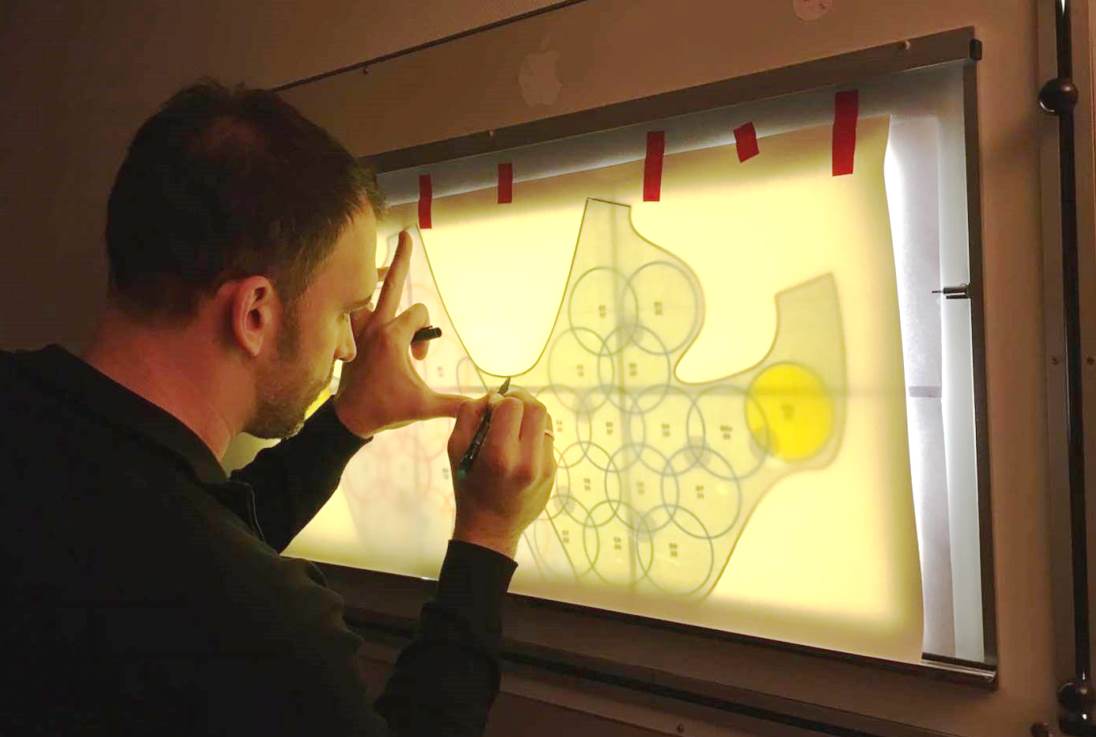
The scientists are developing a vest, or waistcoat of sorts, that features a set of 32 radio frequency coils sewn into the fabric. These eight-centimetre-long (3.1-in) loops hook up to a radio receiver unit by way of coaxial cables, while motion sensors are also built into the vest so the system can cancel out breathing movements in the chest that can distort the image.
"In our case, the woman lies face up and the coils should be flexible enough to be put on like a waistcoat," explains Laistler. "An essential aspect of the project is that lying on the back results in the breast being flattened, which means a much larger part of it is close to the receiver. In this way, the signal is stronger and the measuring time can be shortened."
The waistcoat is under development for now, although the team has published a trail of papers in the past year in Scientific Reports, the Journal of Magnetic Resonance and PLOS One.

Disclaimer: This site is primarily intended for healthcare professionals. Any content/information on this website does not replace the advice of medical and/or health professionals and should not be construed as medical/diagnostic advice/endorsement or prescription. Use of this site is subject to our terms of use, privacy policy, advertisement policy. © 2020 Minerva Medical Treatment Pvt Ltd