- Home
- Editorial
- News
- Practice Guidelines
- Anesthesiology Guidelines
- Cancer Guidelines
- Cardiac Sciences Guidelines
- Critical Care Guidelines
- Dentistry Guidelines
- Dermatology Guidelines
- Diabetes and Endo Guidelines
- Diagnostics Guidelines
- ENT Guidelines
- Featured Practice Guidelines
- Gastroenterology Guidelines
- Geriatrics Guidelines
- Medicine Guidelines
- Nephrology Guidelines
- Neurosciences Guidelines
- Obs and Gynae Guidelines
- Ophthalmology Guidelines
- Orthopaedics Guidelines
- Paediatrics Guidelines
- Psychiatry Guidelines
- Pulmonology Guidelines
- Radiology Guidelines
- Surgery Guidelines
- Urology Guidelines
Craniopagus twins: British doctors separated skulls, brains and blood vessels of conjoined twins

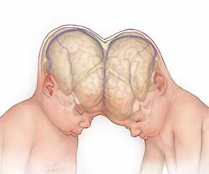
LONDON - Doctors at a British hospital separated skulls, brains and blood vessels of conjoined twins after a 50 hours-long surgery. The operation was performed on the two-year-old Craniopagus twins joined at the head.
The highly complex surgery involved multiple operations on twins, who were born in Pakistan in January 2017 with a condition known as “craniopagus” in which the girls’ skulls and parts of their brains were joined and intertwined.
“Craniopagus is an exceptionally rare and complex condition,” said David J. Dunaway, who co-led the surgical team that treated the twins. The operation, conducted in February, was the most complex such separation his team had performed to date, he said.
Having twins joined at the head with fused skulls and separate bodies occur in less than one in a million births, while having the connection extend into the brain tissue is rarer still. Around 50 sets of craniopagus twins are estimated to be born around the world every year, of which only around 15 are thought to survive beyond the first 30 days of life.
Dunaway said this separation was helped by state-of-the-art technology including virtual reality, advanced imaging, and three-dimensional rapid prototyping - allowing the surgeons to use images of the girls’ brains and blood vessels to plan and practice the surgery in advance to minimize complications.
The procedures took place at London’s Great Ormond Street hospital, with the girls well enough to be discharged from hospital four months later on July 1.
“These cutting-edge scientific techniques greatly increased the chance of success for Safa and Marwa. Their brains were more intertwined than the previous sets of craniopagus twins making it the most complicated separation to date,” the Great Ormond Street team said in a statement.
Five months after their final operation, Safa and Marwa are making slow but steady progress, the doctors said, adding that “a further period of recuperation and rehabilitation is essential to maximize their recovery”.

Disclaimer: This site is primarily intended for healthcare professionals. Any content/information on this website does not replace the advice of medical and/or health professionals and should not be construed as medical/diagnostic advice/endorsement or prescription. Use of this site is subject to our terms of use, privacy policy, advertisement policy. © 2020 Minerva Medical Treatment Pvt Ltd