- Home
- Editorial
- News
- Practice Guidelines
- Anesthesiology Guidelines
- Cancer Guidelines
- Cardiac Sciences Guidelines
- Critical Care Guidelines
- Dentistry Guidelines
- Dermatology Guidelines
- Diabetes and Endo Guidelines
- Diagnostics Guidelines
- ENT Guidelines
- Featured Practice Guidelines
- Gastroenterology Guidelines
- Geriatrics Guidelines
- Medicine Guidelines
- Nephrology Guidelines
- Neurosciences Guidelines
- Obs and Gynae Guidelines
- Ophthalmology Guidelines
- Orthopaedics Guidelines
- Paediatrics Guidelines
- Psychiatry Guidelines
- Pulmonology Guidelines
- Radiology Guidelines
- Surgery Guidelines
- Urology Guidelines
Artificial Intelligence (AI) enhances diagnostic accuracy of breast mammography, finds study

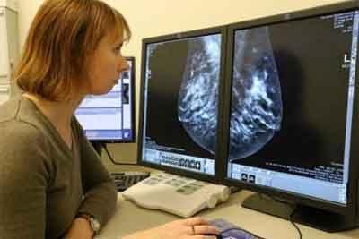
Artificial Intelligence (AI) enhances the diagnostic accuracy of breast mammography and may reduce the chances of missed diagnosis of breast cancer, according to a recent study published in Radiology.
A team of researchers from IBM research trained a combined machine-learning and deep-learning model that identified cancer in nearly half of women who had an initial negative mammogram interpretation but were later found to have biopsy-proven cancer within a year.
Breast cancer is one of the increasing threats that affect millions of people's life. This requires timely and precise breast cancer diagnosis. Artificial Intelligence specifically deep learning has demonstrated remarkable performance in image recognition tasks. The technology has myriad applications in the medical image analysis field, propelling it forward at a rapid pace. Computational models on the basis of deep neural networks are increasingly used to analyze health care data. However, the efficacy of traditional computational models in radiology is a matter of debate.
The study was conducted to evaluate the accuracy and efficiency of a combined machine and deep learning approach for early breast cancer detection applied to a linked set of digital mammography images and electronic health records.
Ayelet Akselrod-Ballin et al from the Department of Healthcare Informatics, University of Haifa Campus, Mount Carmel conducted a retrospective study where 52 936 images were collected in 13 234 women who underwent at least one mammogram between 2013 and 2017, and who had health records for at least 1 year before undergoing mammography.
The algorithm was trained on 9611 mammograms and health records of women to make two breast cancer predictions: to predict biopsy malignancy and to differentiate normal from abnormal screening examinations. The study estimated the association of features with outcomes by using t-test and Fisher exact test. The model comparisons were performed with a 95% confidence interval (CI) or by using the DeLong test.
Key finding of the study
- The researchers validated the resulting algorithm in 1055 women and tested in 2548 women (mean age, 55 years ± 10 ).
- In the test set, the algorithm identified 34 of 71 (48%) false-negative findings on mammograms.
- For the malignancy prediction objective, the algorithm obtained an area under the receiver operating characteristic curve (AUC) of 0.91, with a specificity of 77.3% at a sensitivity of 87%.
- When trained on clinical data alone, the model performed significantly better than the Gail model.
The algorithm, which combined machine-learning and deep-learning approaches, can be applied to assess breast cancer at a level comparable to radiologists and has the potential to substantially reduce missed diagnoses of breast cancer, wrote the researchers.
For more reference, click on the link
https://doi.org/10.1148/radiol.2019182622

Disclaimer: This site is primarily intended for healthcare professionals. Any content/information on this website does not replace the advice of medical and/or health professionals and should not be construed as medical/diagnostic advice/endorsement or prescription. Use of this site is subject to our terms of use, privacy policy, advertisement policy. © 2020 Minerva Medical Treatment Pvt Ltd