- Home
- Editorial
- News
- Practice Guidelines
- Anesthesiology Guidelines
- Cancer Guidelines
- Cardiac Sciences Guidelines
- Critical Care Guidelines
- Dentistry Guidelines
- Dermatology Guidelines
- Diabetes and Endo Guidelines
- Diagnostics Guidelines
- ENT Guidelines
- Featured Practice Guidelines
- Gastroenterology Guidelines
- Geriatrics Guidelines
- Medicine Guidelines
- Nephrology Guidelines
- Neurosciences Guidelines
- Obs and Gynae Guidelines
- Ophthalmology Guidelines
- Orthopaedics Guidelines
- Paediatrics Guidelines
- Psychiatry Guidelines
- Pulmonology Guidelines
- Radiology Guidelines
- Surgery Guidelines
- Urology Guidelines
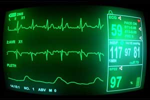
AHA updates practice standards for Inpatient ECG Monitoring

Sandau KE et al. formalized Updates to the American Heart Association (AHA) Scientific Statement on Practice Standards for Electrocardiographic (ECG) Monitoring in Hospital Settings originally published in 2004. They were entrusted for the job by the American Heart Association.The committee also included experts from general cardiology, electrophysiology (adult and pediatric), and interventional cardiology, as well as a hospitalist and experts in alarm management. The statement was published online by Circulation.
One significant difference from the 2004 practice standards was a downgrading of the recommendation for continuous ST-segment monitoring for certain patients at risk of myocardial ischemia not because we doubt the potential for this technology but because recent literature demonstrates a very serious problem with false and nonactionable alarm signals.
The recommendations are graded according to their level of evidence support, but the authors noted that many of the recommendations are based on limited data.The salient points of recommendations are:
- Appropriate electrode placement and lead selection are paramount to ensure proper continuous rhythm monitoring. In adults, lead V1 is commonly selected for arrhythmia monitoring due to its ability to help discriminate between ventricular tachycardia (VT) and aberrancy. In children, lead II is commonly selected as the primary lead due to supraventricular arrhythmias being more common than ventricular arrhythmias and P waves being more easily seen in the inferior leads.
- It is reasonable (Class IIa) to perform ST-segment monitoring in patients during the early phase of an acute coronary syndrome; post-myocardial infarction (MI) without revascularization or with residual ischemic lesions until there is no evidence of ongoing modifiable ischemia or hemodynamic/electrical instability; newly diagnosed left main coronary artery lesion until revascularized; after nonurgent percutaneous coronary intervention with complications; and intraoperatively during open heart surgery. Continuous ECG monitoring of the ST-segment might be considered (Class IIb) in patients post-MI with revascularization of all lesions; stress cardiomyopathy; therapeutic hypothermia; post-open heart surgery; acute decompensated heart failure; and in stroke in patients with increased risk for cardiac events.
- The ECG lead with the longest T wave should be selected when monitoring the QT interval, and leads with U waves should be avoided. It is recommended (Class I) that patients, regardless of their risk for torsades de pointes (TdP) who are started on medications with an established or possible risk of TdP, be monitored with continuous ECG.
- Patients undergoing targeted temperature management should remain on continuous ECG monitoring until the temperature is normalized, the QTc interval is within the normal range, and there is no evidence of QT-related arrhythmias (Class I). It is also recommended (Class I) that patients with inherited long-QT who present with unstable ventricular arrhythmias or have medically or metabolically induced QTc prolongation to have continuous ECG monitoring. Patients with significant hypokalemia or hypomagnesemia with other risk factors for TdP should also be monitored (Class I). Finally, patients who suffered from a drug overdose with a drug having known TdP risk, or with a drug overdose of unknown type should have continuous ECG monitoring (Class I).
- Patients resuscitated from cardiac arrest or unstable VT have a high risk of recurrent arrhythmia and should have continuous ECG monitoring. These patients should remain on monitoring until implantable cardioverter-defibrillator (ICD) implantation (Class I). Patients with premature ventricular contractions and nonsustained VT who do not have other indications for monitoring can be considered for continuous ECG monitoring (Class IIb). Patients with an ICD and shocks requiring hospital admission should be maintained on continuous ECG monitoring for the duration of the related hospitalization (Class I).
- For unidentified supraventricular arrhythmias, continuous ECG will aid in diagnosis and management. Patients admitted for new onset or recurrent atrial arrhythmias, hemodynamically unstable or symptomatic atrial arrhythmias, or if rate control for arrhythmia is deemed necessary, should be on continuous ECG monitoring (Class I). It is recommended that patients with symptomatic or asymptomatic significant sinus bradycardia, or symptomatic second-degree atrioventricular (AV) block or third-degree AV block have continuous ECG monitoring. Hemodynamically unstable arrhythmic conditions such as Wolff-Parkinson-White syndrome should also be monitored on continuous ECG until appropriate therapy is delivered.
- Patients with mechanical circulatory support devices (e.g., total artificial heart, extracorporeal life support, ventricular assist device, intra-aortic balloon pump) should have continuous ECG monitoring (Class I). The goal of continuous ECG monitoring in these patients is to detect atrial and ventricular arrhythmias and when applicable, ischemia. Patients with mechanical circulatory support who are admitted to a rehabilitation facility are not recommended to have continuous ECG monitoring (Class III).
- Patients with transcatheter interventions also should be monitored with continuous ECG. Patients post-transcatheter aortic valve replacement (TAVR) should be monitored for at least 3 days post-procedure to detect AV block, arrhythmia, and development of bundle branch block. Following the transcatheter closure of an atrial septal defect or ventricular septal defect, continuous ECG monitoring is also recommended to detect bundle branch block, arrhythmia, and AV block (Class I).
- Continuous ECG monitoring is recommended for patients with syncope of unknown etiology meeting admission criteria. Patients with stroke may have continuous ECG monitoring for 24-48 hours or for longer time periods if cryptogenic stroke and occult atrial fibrillation is suspected. Patients following noncardiac thoracic surgery should be considered for continuous ECG monitoring due to the risk of atrial fibrillation in this patient population (Class IIa).
- Adequate education for medical staff is critical for ECG monitoring and correct interpretation of waveforms and data. Education goals should include the ability to: identify ST changes consistent with ischemia; accurately measure a QTc interval; identify common arrhythmias; and enact the appropriate response to an identified ECG abnormality. Addressing proper alarm management and alarm fatigue is an important aspect of continuous ECG monitoring and is best performed through an interdisciplinary committee.
It is important to note that proper documentation of ECG monitoring is a must which will vary based on the ability to interface with current electronic medical record systems. If no mechanism is available to transfer ECG strips to the electronic medical records, it is critical that paper ECG strips be printed and maintained in a paper chart and scanned into the electronic health record. For ST-segment and QTc monitoring, relevant parameters should be documented at baseline and then at least every 8-12 hours.
For further details log on to :
http://circ.ahajournals.org/content/early/2017/10/03/CIR.0000000000000527

Disclaimer: This site is primarily intended for healthcare professionals. Any content/information on this website does not replace the advice of medical and/or health professionals and should not be construed as medical/diagnostic advice/endorsement or prescription. Use of this site is subject to our terms of use, privacy policy, advertisement policy. © 2020 Minerva Medical Treatment Pvt Ltd