- Home
- Editorial
- News
- Practice Guidelines
- Anesthesiology Guidelines
- Cancer Guidelines
- Cardiac Sciences Guidelines
- Critical Care Guidelines
- Dentistry Guidelines
- Dermatology Guidelines
- Diabetes and Endo Guidelines
- Diagnostics Guidelines
- ENT Guidelines
- Featured Practice Guidelines
- Gastroenterology Guidelines
- Geriatrics Guidelines
- Medicine Guidelines
- Nephrology Guidelines
- Neurosciences Guidelines
- Obs and Gynae Guidelines
- Ophthalmology Guidelines
- Orthopaedics Guidelines
- Paediatrics Guidelines
- Psychiatry Guidelines
- Pulmonology Guidelines
- Radiology Guidelines
- Surgery Guidelines
- Urology Guidelines
A classical case of Zollinger-Ellison syndrome

Dr Ali Alshati department of Internal Medicine, Maricopa Integrated Health System, Creighton University, Phoenix, AZ, USA, and colleagues have reported a classical case of Zollinger-Ellison syndrome. The case has appeared in the journal Gastrointestinal Endoscopy.
Zollinger-Ellison syndrome is a rare condition in which one or more tumours form in the pancreas or the upper part of small intestine (duodenum). These tumours, called gastrinomas, secrete large amounts of the hormone gastrin, which causes the stomach to produce too much acid. The excess acid then leads to peptic ulcers, as well as to diarrhoea and other symptoms.
A 64-year-old man presented with watery diarrhoea and 40-pound weight loss. He has a history of gastroesophageal reflux disease and esophagitis seen in upper endoscopy done previously. His epigastric discomfort and heartburn symptoms were getting worse with eating and were poorly responsive a daily dose of 40 mg oral omeprazole. The diarrhea was not improving in response to fasting. He denied dysphagia or odynophagia.
Laboratory workup showed fasting gastrin level 669 pg/mL, serum chromogranin A 100 ng/l and gastric PH at 1. A CT scan with contrast of the abdomen and pelvis showed a normal pancreas. However, it revealed multiple, solid with cystic component liver lesions that are compatible with metastatic disease. CT-guided core-needle liver biopsy revealed metastatic well-differentiated neuroendocrine carcinoma/carcinoid tumor. The primary tumor was undetermined.
Upper endoscopy demonstrated diffuse erosive inflammation of the second part of the duodenum. Closer endoscopic look underwater magnification showed the hypertrophied parietal cells. Duodenal biopsy was consistent with chronic peptic duodenitis with no evidence of dysplasia or malignancy.
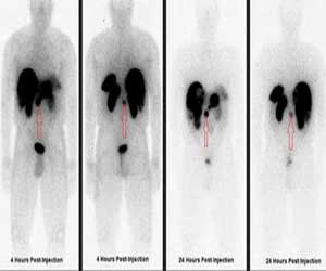
EUS demonstrated a single, well-defined 2x2, round, hypoechoic, heterogeneous solid mass in the head of the pancreas. The outer margin of the mass was slightly irregular. EUS also revealed a gastric wall thickening. Beside the physiologic uptake in the kidneys and the spleen, the pentetreotide scan demonstrated intense focal uptake in a 2.3 cm soft tissue related to the ventral surface of the pancreatic head, consistent with malignancy.
It also revealed multiple radiotracer avid liver metastases. The patient was treated with long-acting octreotide 30 mg and experienced relief of symptoms.
For more details click on the link: DOI: https://doi.org/10.1016/j.gie.2019.01.026

Disclaimer: This site is primarily intended for healthcare professionals. Any content/information on this website does not replace the advice of medical and/or health professionals and should not be construed as medical/diagnostic advice/endorsement or prescription. Use of this site is subject to our terms of use, privacy policy, advertisement policy. © 2020 Minerva Medical Treatment Pvt Ltd